Rare diagnosis: When general symptoms hide a deeper issue
Nausea, difficulty swallowing, headache: Such general symptoms can also hide a rare disease. Some important differential diagnoses.
Warning signs of rare diseases in radiology
Swallowing difficulties
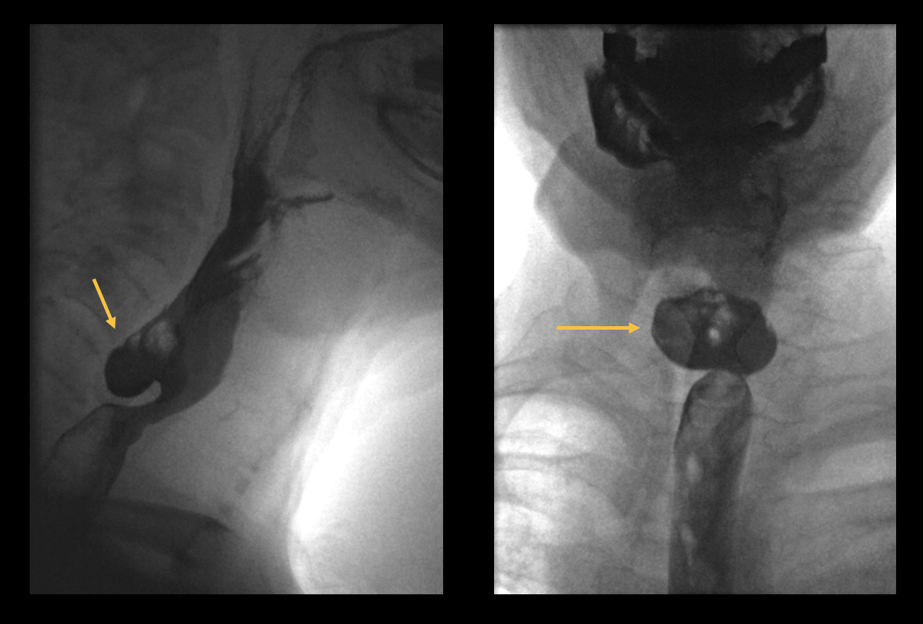
Swallowing difficulties are relatively common in old age. They can be short-termed, but can also last longer. If the symptoms persist for a long time, a gastroscopy should be performed. If this does not reveal anything, a porridge swallowing test can also be helpful. The 65-year-old patient (see illustrations), for example, has a rare finding: a Zenker's diverticulum.
Figure 1: Hypopharyngo-oesophagography with barium-containing contrast medium in sagittal and coronal view. A dorsal protrusion of the posterior oesophageal wall at the level of HWK5/6 is shown.
Tinnitus
Tinnitus (ringing in the ears) is not initially a disease in its own right, but a symptom of various disorders. In the beginning, there is often damage and illness in the ear itself, for example due to inflammation or exposure to strong noise. If the symptoms worsen, it is advisable to have a magnetic resonance imaging (MRI).
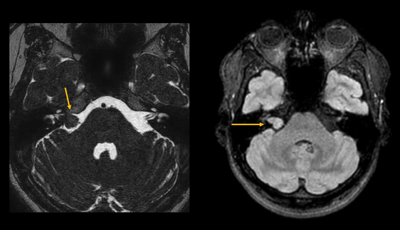
A 47-year-old female patient (see pictures) was diagnosed with an acoustic neuroma on the right side, which led to a hearing loss.
Figure 2: MRI of the posterior fossa in T2 and T1 weighting. This shows a T2-W isointense mass in the internal acoustic meatus, consistent with an acoustic neuroma.
Nausea / vomiting
Patients with persistent nausea and vomiting should always be examined radiologically if the symptoms persist for more than two weeks and the cause is unclear, because a rare disease may be present.
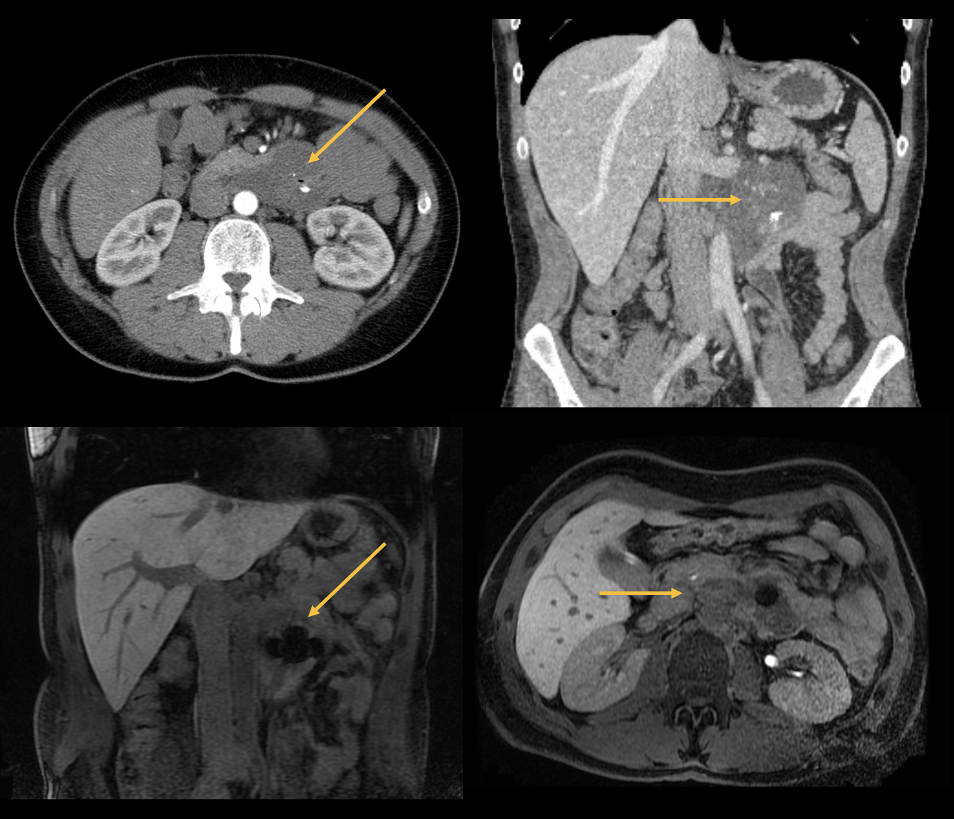
Mr H. complained of non-specific complaints such as bloating, abdominal pain, nausea and anaemia. Imaging (see images) revealed a gastrointestinal stromal tumour (GIST).
GIST is a semi-malignant tumour of the gastrointestinal tract (especially the stomach and small intestine), which can metastasise but is usually associated with a good prognosis. The tumour usually occurs in middle age (40-60) and is treated surgically. In case of metastases, adjuvant immunotherapy with a protein kinase inhibitor is given.
Figure 3: Axial and coronal CT and MRI images. GIST originating from the pars horizontalis duodeni, located ventral to the left renal vein, and adjacent to the pancreatic tail. Central susceptibility artefacts with air pockets and calcifications.
Headache
Headaches are one of the most common pain syndromes that doctors are confronted with in everyday clinical practice. A distinction is made between primary (e.g. migraine) and secondary (e.g. after craniocerebral trauma or caused by infections) headaches.
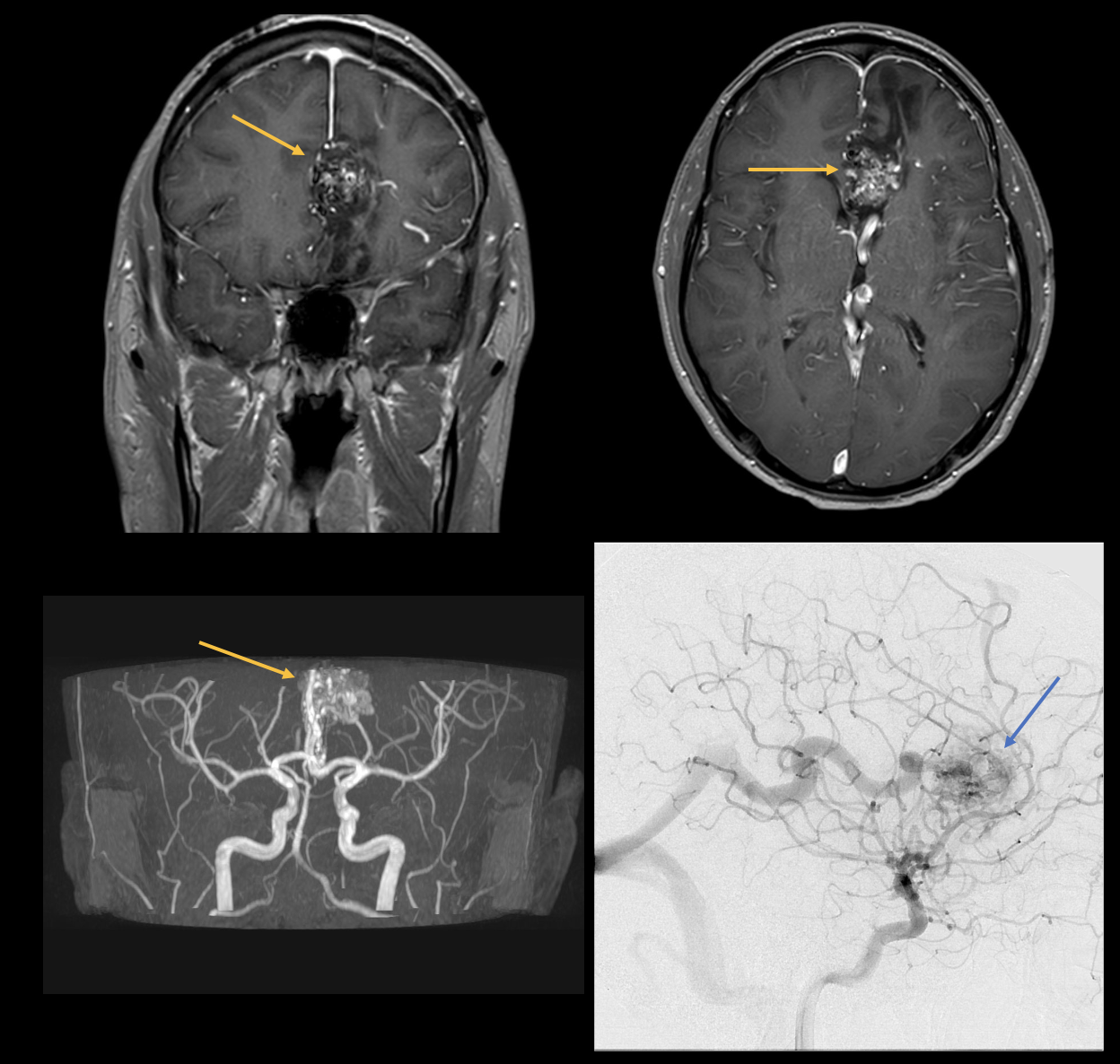
The following pictures show a patient who was admitted to the clinic with severe headache and nausea. MRI and angiography show an arteriovenous malformation (AVM).
An AVM is a congenital vascular malformation with tangles of abnormal blood vessels (nidus) between cerebral arteries and veins without an intermediate capillary bed. Arteriovenous malformations can remain asymptomatic throughout life. However, they often manifest with headaches between the ages of 20 and 40. Intracranial haemorrhages occur in about 50 to 60 % of patients, and epileptic seizures and focal neurological deficits in about 25 % of those affected. The classic therapy is surgical resection, although endovascular treatment is also increasingly being used.
Figure 4: Axial and coronal MRI images, as well as digital subtraction angiography (DSA) showing the tangled connections between the arteries and veins. The nidus is marked by the arrows.
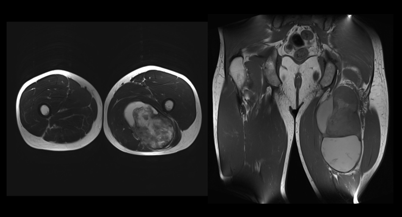
Muscle swelling
It is not uncommon for muscle swelling to occur in the upper or lower leg due to deep vein thrombosis (DVT). These are always ruled out first. If DVT is not confirmed, imaging should be ordered to rule out other causes.
In Mr. K.'s case, magnetic resonance imaging showed a growth originating in the left thigh muscle, which turned out to be a sarcoma. Sarcomas are malignant and highly aggressive tumours of mesenchymal origin. Sarcomas are much rarer overall than carcinomas. They make up about 1% of malignant neoplasms, but they can metastasise and have a lethal course. The primary therapy in this case would be total resection. If the femur is affected, amputation of the limb would have to be considered.
Figure 5: Axial and coronal MRI images of the thighs on both sides. This showed a heterogeneous cystic and partly solid mass originating from the biceps femoris caput breve muscle on the left.
Rare Disease Day